
Ultrasound Scans – How Do They Work?
Ultrasound scanning is one of the most widely used diagnostic imaging techniques in modern medicine, supporting assessment across musculoskeletal, abdominal, vascular, and obstetric care [1]. Unlike X-rays or CT scans, ultrasound uses sound waves rather than ionising radiation, making it a safe, non-invasive, and repeatable imaging option across a wide range of clinical contexts [1].
At its core, ultrasound provides real-time visualisation of soft tissues, organs, blood flow, and dynamic movement [1]. This allows clinicians to not only identify structural changes but also assess function, motion, and response to applied pressure.
Understanding how ultrasound scans work – and what they can and cannot show – helps patients appreciate why they are often chosen as a first-line investigation and how they support accurate diagnosis and personalised care.
Understanding the Basic Principles of Ultrasound Scanning
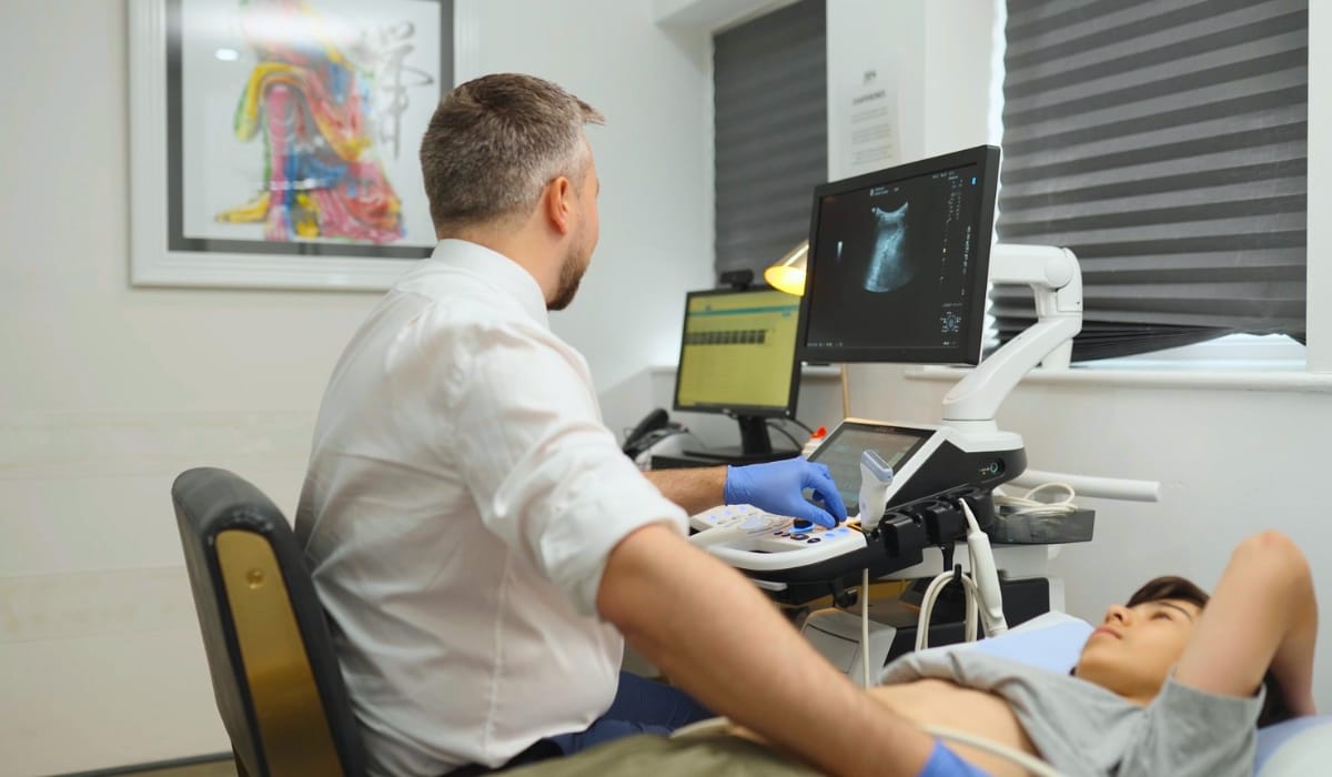
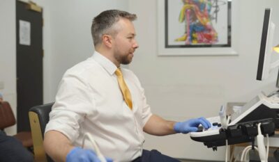
Ultrasound imaging is based on the transmission and reflection of high-frequency sound waves, typically in the range of 2–15 megahertz [2]. These sound waves are emitted from a handheld device called a transducer, which is placed on the skin over the area being examined [1][2].
As the pulsed sound waves travel through the body, they interact with tissues of different densities. Some waves pass through soft tissues, while others are reflected when they encounter boundaries between structures, such as muscle and tendon or fluid and solid tissue [1].
The returning echoes are detected by the transducer and converted into electrical signals, which are then processed by a computer to create a scaled internal image [1]. The time it takes for echoes to return, along with their intensity, determines the depth and appearance of structures on the screen.

Because sound waves travel differently through various tissues, ultrasound is particularly well suited to visualising soft tissue structures, fluid-filled spaces, and dynamic movement in real time [3].
Exploring Advances and What Ultrasound Scanning Can Visualise
Ultrasound excels at imaging soft tissues that are less visible on plain X-ray imaging. This includes muscles, tendons, ligaments, bursae, nerves, blood vessels, and many internal organs [4][5]. In musculoskeletal medicine, ultrasound is frequently used to assess tendon tears, muscle injuries, ligament sprains, joint effusions, and inflammatory changes.
Modern ultrasound systems even go beyond the idea of static grey scale images, offering high-resolution visualisation of soft tissues, joints, and dynamic movement in real time. This allows clinicians to evaluate structures as you move or as gentle pressure is applied, linking what is seen on the screen directly to the symptoms you feel [6][7].
High-frequency musculoskeletal ultrasound has shown excellent diagnostic accuracy for tendon injuries, with studies reporting accuracy rates above 90% for certain tendon ruptures (~93% for flexor tendons and 100% for extensor tendons) [8]. It can reveal subtle structural changes such as early tendon degeneration, small tears, and fluid accumulation that may not be visible on X-ray and may only be picked up on more advanced imaging at a later stage [8]. Dynamic ultrasound assessment has also been used to evaluate how structures like the meniscus move during joint motion, helping to identify early functional changes that guide timely investigation and treatment [8][9][10].
This ability to assess both structure and movement in a single appointment supports quicker, better-targeted clinical decision-making and often reduces the need for more complex scans [8][9][10]. As well as this, advanced imaging modes such as extended field-of-view and 3D-style reconstructions can improve anatomical orientation in larger regions, such as the shoulder or thigh, giving a clearer overview of the area being assessed. For patients, these technological advances translate into detailed, radiation-free imaging that can be integrated seamlessly into a routine consultation.
Comparing Ultrasound Scanning With Other Imaging Modalities
Each imaging technique obviously has its own distinct strengths and limitations, and ultrasound is often chosen because of its accessibility, safety profile, and ability to provide real-time information. Unlike X-rays and CT scans, ultrasound does not use ionising radiation. This makes it suitable for repeated use and for populations where radiation exposure is a concern [12]. Compared with MRI, ultrasound is faster, more cost-effective, and allows dynamic assessment (assessing structures as they move). However, MRI may be superior for deep structures or complex joint pathology, particularly where bone marrow or cartilage detail is required [12].
Large-scale analyses show ultrasound has diagnostic accuracy comparable to MRI for many soft tissue conditions. For example, a meta-analysis found ultrasound detected full-thickness rotator cuff tears with 91% sensitivity and 95% specificity – matching MRI performance – making it the preferred first-line tool, with MRI reserved for complex cases [13].
Assessing Accuracy and Reliability in Clinical Practice
The diagnostic accuracy of ultrasound depends on both the quality of equipment and the expertise of the clinician performing and interpreting the scan. When conducted by specialist practitioners, ultrasound demonstrates high reliability across multiple applications. For example, in musculoskeletal imaging, systematic reviews report diagnostic accuracy rates exceeding 85-90% for common conditions such as tendon tears, muscle injuries, and joint effusions [7][9][14].
A 2023 review of point-of-care ultrasound in musculoskeletal medicine found that ultrasound-guided diagnosis altered clinical management in over 60% of cases, highlighting its potential impact on informed treatment decision-making [15]. Importantly, ultrasound’s ability to correlate imaging findings directly with a patient’s symptoms – through guided movement and palpation during scanning – enhances clinical relevance beyond static images alone.
Considering Safety and the Patient Experience
Ultrasound is widely regarded as one of the safest imaging modalities available. It does not involve radiation, contrast agents, or invasive procedures, and adverse effects are exceedingly rare. From a patient perspective, ultrasound scans are generally well tolerated. The procedure typically lasts between 15 and 30 minutes, involves minimal discomfort, and results can often be discussed immediately [16] [17].
This immediacy supports shared decision-making, allowing patients to better understand their condition and the rationale behind recommended treatments. Research suggests that real-time explanation of imaging findings improves patient engagement and satisfaction with care [18]. Because ultrasound is portable and flexible, it can be integrated seamlessly into clinical assessments, reducing delays between investigation and intervention [2] [18].
Supporting Ultrasound-Guided Interventions
Beyond diagnosis, ultrasound plays a growing role in guiding interventional procedures. Ultrasound guidance improves the accuracy of injections by allowing clinicians to visualise needle placement in real time. Studies consistently show that ultrasound-guided injections achieve higher accuracy rates than landmark-guided techniques. For example, a systematic review found that ultrasound-guided hip joint injections achieved 100% accuracy compared with 72% using anatomical landmarks alone [19].
Improved accuracy is associated with better clinical outcomes, reduced complication rates, and more consistent symptom relief. This is particularly relevant for injections involving small joints, tendons, or peri-neural structures.
Integrating Ultrasound Into Personalised Care Pathways
Ultrasound is most effective when used as part of an integrated clinical assessment rather than in isolation. Imaging findings must be interpreted in the context of symptoms, physical examination, activity demands, and medical history [16]. By combining ultrasound with functional assessment, clinicians can identify not only structural changes but also biomechanical contributors to pain or dysfunction. This supports more tailored rehabilitation, targeted interventions, and realistic expectations around recovery [16].
In many cases, ultrasound findings can guide conservative management strategies, monitor progress over time, and reduce unnecessary referrals for more invasive investigations or procedures.
Applying Ultrasound Scanning Expertise at The Health Suite
Whether used to clarify a diagnosis, guide treatment decisions, or support ultrasound-guided interventions, imaging is tailored to your symptoms, goals, and long-term health needs. The focus is not simply on identifying abnormalities, but on understanding how findings relate to movement, function, and recovery.
At The Health Suite Leicester, ultrasound scanning is delivered within a clinician-led, patient-centred framework by experienced sonographers who integrate real-time imaging findings directly into your clinical assessment and personalised care plan. Our radiation-free, non-invasive approach provides clear insights for better health outcomes, whether you’re seeking early diagnosis, treatment guidance, or progress monitoring across musculoskeletal conditions.
This safe, versatile technology offers zero ionising radiation risk – ideal for repeated use, pregnancy monitoring, and all age groups – while advanced 3D/4D capabilities provide enhanced anatomical detail when clinically indicated. Whether investigating sports injuries, chronic pain, or preventive health checks, our private scans deliver fast appointments, specialist insights, and tailored recommendations to support your movement, function, and long-term wellbeing.
Book a Ultrasound Scan in Leicester
If you are experiencing unexplained symptoms, discomfort, or simply need reassurance, an ultrasound scan can provide quick, non-invasive insight into what may be going on inside the body. At The Health Suite Leicester, we offer fast access to diagnostic imaging in a comfortable, patient-focused setting.
Our ultrasound services are designed to support early diagnosis and informed clinical decision-making, helping to guide the most appropriate next steps in your care.

Adult Ultrasound Scanning
Private adult ultrasound scanning in Leicester. Get safe, radiation-free imaging and same-day diagnostic results from expert consultant radiologists.
Paediatric Ultrasound Scan Services
Private paediatric ultrasound scans in Leicester. Safe, radiation-free imaging for infants and children under expert consultant care.
References
Have a query about Ultrasound Scans – How Do They Work??
We recognise that getting the healthcare assistance you need can be difficult. So if you have a query, feel free to contact us and one of our treatment co-ordinators will be happy to help. We aim to reply to all queries within 24 hours (Mon – Fri).