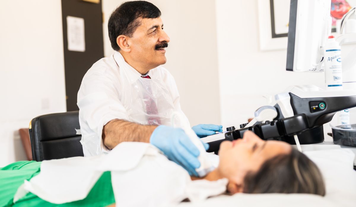
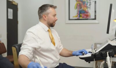
Adult Ultrasound Scanning in Leicester
Clear Insights, Better Health – Expert Ultrasound Scanning at The Health Suite Leicester
Ultrasound scanning, also known as sonography, is a non-invasive and radiation-free imaging technique used to examine internal body structures in real time. It works by emitting high-frequency sound waves through a device called a transducer, which sends the waves into the body and captures the echoes that bounce back from tissues, organs, and fluids. A computer then processes these echoes into detailed, live images, allowing healthcare professionals to assess various medical conditions. This service is suitable for those aged 18 and over, if you want to book a scan for those aged under 18, please click here: Paediatric Ultrasound Scan Services.
Advantages of Ultrasound Scanning
- Safe and Radiation-Free – Ultrasound is a non-invasive imaging technique that does not use ionising radiation, making it a safe choice for patients of all ages. Unlike X-rays or CT scans, ultrasound poses no radiation risk, making it suitable for repeated use when necessary, and ideal for monitoring both maternal and fetal health throughout pregnancy.
- Versatile and Widely Used – Ultrasound is a highly versatile tool that can be used for a variety of purposes. It is especially effective in imaging soft tissues, such as muscles, tendons, and organs, and can help assess the health of internal structures like the heart, liver, kidneys, and reproductive organs. Additionally, ultrasound is used to guide medical procedures, such as biopsies, injections, or fluid drainage, ensuring precision and reducing complications.
- Real-Time Imaging – One of the key benefits of ultrasound is its ability to provide real-time imaging, allowing healthcare professionals to visualise internal structures dynamically. This capability is especially useful in assessing blood flow, organ movement, or the condition of muscles and joints, and helps guide immediate decision-making during medical procedures. The real-time aspect also enables immediate feedback, facilitating quicker diagnoses and treatment plans.
Private Ultrasound Scanning at The Health Suite Leicester
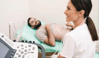
At The Health Suite Leicester, we offer expert-led private ultrasound services in a comfortable and professional setting. Our scans are used for a variety of purposes, including abdominal and pelvic health assessments, musculoskeletal conditions, and vascular imaging. With fast appointments, expert sonographers, and comprehensive reports, we provide high-quality diagnostic imaging to support accurate and timely medical decision-making. Whether for a routine health check or investigating specific symptoms, ultrasound scanning is an essential tool for early detection, diagnosis, and treatment planning.
Watch to learn more about this treatment
Watch expert videos on your treatment to understand the benefits, process, and what to expect before your consultation.
Abdominal Ultrasound Scans | How They Work & Why They’re Important | Dr. Deepak Kathuria | Leicester
Joint Ultrasound Scans Explained | Benefits & How They Work | Dr. Deepak Kathuria | The Health Suite
What Is a Scrotal Ultrasound? | Safe & Quick Test for Testicular Health | The Health Suite Leicester
What we treat
At The Health Suite Leicester, our private medical clinic offers expert care whenever you need it. Our team of experienced GPs and healthcare professionals provide personalised diagnosis and treatment for a wide range of medical conditions, ensuring high-quality, professional care in a comfortable setting.
Click below to view useful info on a few of the common conditions we treat:
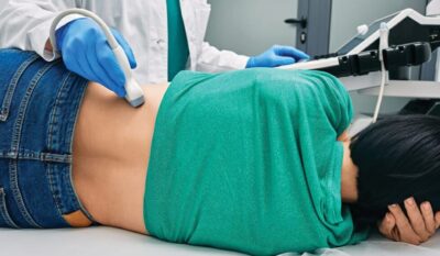
Abdominal Organ Assessment
Examine internal digestive organs non-invasively. We scan the liver, gallbladder, pancreas, and kidneys to pinpoint the root causes of persistent abdominal pain, bloating, or abnormal laboratory test results.
Pelvic Health Imaging
Identify structural abnormalities in pelvic organs. Our targeted scans evaluate the bladder, uterus, ovaries, or prostate to guide clinical care for unexplained pelvic discomfort and sudden urinary changes.
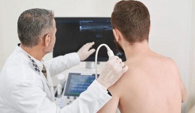
Musculoskeletal & Soft Tissue Evaluation
Diagnose muscle, tendon, and ligament damage accurately. High-frequency scans evaluate acute soft-tissue inflammation, sports-related tears, painful joint fluid build-up, and unexplained physical lumps.
Vascular & Circulatory Assessments
Track deep blood flow patterns. Using advanced Doppler ultrasound technology, we assess arteries and veins to identify circulation irregularities driving cold extremities, pain, or limb swelling.
Diagnostic Support for Unexplained Symptoms
Clear up diagnostic uncertainties safely. When persistent discomfort, swelling, or abnormal lab tests leave you without clear answers, real-time scanning provides vital clarity to guide your treatment.
many more
Our clinicians manage a broad spectrum of conditions, and individual assessment allows us to tailor care beyond the examples listed. We encourage you to book a consultation to discuss your symptoms and appropriate treatment options.
Symptom checker
If you are experiencing any of these symptoms, please book an appointment with us. Symptoms can vary from person to person and from condition to condition. A proper diagnosis can only be made through a thorough medical examination.
Our clinicians will carry out a full assessment to ensure an accurate diagnosis and appropriate care.
Do you have persistent abdominal pain or bloating?
Abdominal discomfort, bloating, or unexplained pain may be due to problems with organs like the liver, gallbladder, or kidneys. Ultrasound helps visualise these structures safely and non‑invasively to find the cause.
Have you noticed lumps, swelling, or soft‑tissue changes?
Ultrasound scanning can assess lumps or swellings in muscles, soft tissues, or glands, helping determine if further investigation or treatment is needed.
Do you have unusual urinary or pelvic symptoms?
Symptoms like pain during urination, pelvic discomfort, or abnormal bleeding may relate to bladder, prostate, or reproductive organ issues—and an ultrasound can examine these safely.
Are you experiencing joint pain, weakness, or tendon issues?
Ultrasound is often used in musculoskeletal assessments to look at joints, tendons, ligaments, and soft tissue structures, helping diagnose tears, inflammation, or injury.
Do you have symptoms linked to blood flow or vascular concerns?
If you notice coldness, colour changes, or swelling in limbs, a vascular ultrasound (like Doppler) can assess arterial or venous blood flow to help diagnose circulation problems.
Are you concerned about organ‑related symptoms without a clear cause?
Unexplained symptoms involving internal organs—such as nausea, back pain, or abnormal blood tests—can be investigated with an ultrasound to give real‑time insights and guide diagnosis.
Why choose The Health Suite for Ultrasound Scanning?
Choose The Health Suite Leicester for expert, personalised healthcare, fast access to specialists, advanced diagnostics and exceptional patient care.
Experienced Consultants
All of our ultrasound clinic doctors are Fellows of The Royal College of Radiologists and have years of experience as consultants in their respective specialist areas. In addition, many of them incorporate both diagnostic and interventional radiology disciplines.
State-of-the-Art Equipment
At The Health Suite Leicester, we use modern, high-quality ultrasound equipment that offers superior image clarity, helping to detect and diagnose medical conditions more effectively.
Patient-Centred Care
We prioritise patient comfort, communication, and education, ensuring that individuals receive personalised attention and understand the procedure and its results thoroughly. Patients are given same-day results, and if required, an onward referral can be facilitated.
Common Q&A about Ultrasound Scanning
Our FAQ section is designed to address common questions you may have, from how our treatments work to what you can expect during and after your session.
Our team is always available to provide additional support if you need more personalised guidance, ensuring that you feel informed and confident every step of the way.
Advice from the experts
Read clear, expert advice from our clinicians, offering trusted advice, clinical insight, and practical guidance to help you understand your treatment and care options.
Ultrasound Scans – How Do They Work?
Ultrasound Scans – Weighing Up the Pros and Cons
How to Prepare for an Abdominal Ultrasound Scan
Meet our Adult Ultrasound Scanning Clinicians
Meet our dedicated team at The Health Suite Leicester, where skilled professionals are committed to providing exceptional care tailored to your medical and wellness needs.

Dr. Rajashri Patil

Dr Jonathan Delf

Professor Arumugam Rajesh

Dr Deepak Kathuria
Treatment options and pricing
Browse and click below to book any of our available services.
Abdomen Ultrasound Scan
Abdomen ultrasound scan is a non-invasive imaging test that uses sound waves to examine organs such as the liver, kidneys, and gallbladder for abnormalities or disease.
An upper abdomen ultrasound scan uses sound waves to examine organs like the liver, gallbladder, pancreas, and kidneys, helping detect abnormalities or conditions.
The upper abdomen ultrasound is ideal for assessing conditions related to the pancreas, liver, kidneys, gall bladder, spleen, and aorta.
Symptoms like abdominal pain, jaundice, unexplained weight loss, and digestive issues may indicate problems in these areas.
It’s particularly useful for evaluating liver diseases, kidney stones, and issues related to the pancreas and gall bladder.
This scan can reveal the size, shape, and any abnormalities in the upper abdominal organs.
It’s effective in detecting cysts, tumours, blockages in the bile ducts, kidney stones, and changes in the aorta.
This ultrasound provides crucial insights into the health and function of these vital organs.
Please note: This scan is not suitable for gynaecological purposes.
A lower abdomen ultrasound scan uses sound waves to examine organs like the bladder, uterus, ovaries, and prostate, helping diagnose issues such as cysts or inflammation.
A lower abdomen ultrasound scan is a safe, non-invasive test that uses sound waves to create detailed images of internal organs, including the bladder, uterus, ovaries, and prostate.
It helps detect conditions such as cysts, inflammation, or structural abnormalities, supporting accurate diagnosis and treatment planning.
Please note: This scan is not suitable for gynaecological purposes.
An upper and lower abdomen ultrasound scan examines both the upper and lower abdominal organs, to detect abnormalities.
This comprehensive scan covers both the upper and lower abdomen, suitable for a range of symptoms affecting the abdomen.
It’s recommended for patients experiencing comprehensive abdominal issues, providing a thorough evaluation of the abdominal region.
It combines the insights of both upper and lower abdomen scans, offering a detailed view of the abdominal organs.
This can help in diagnosing a wide range of conditions affecting these areas.
Please note: This scan is not suitable for gynaecological purposes.
Abdomen and Scrotum Ultrasound Scan
Abdomen and scrotum ultrasound scan is a non-invasive test that uses sound waves to assess abdominal organs and testicular health, helping detect abnormalities early.
An abdomen and scrotum ultrasound scan examines the abdominal organs (liver, kidneys, etc.) and scrotum to detect issues like cysts, hernias, or abnormalities in the testes.
This scan is beneficial for evaluating abdominal issues along with scrotal or testicular problems.
It’s recommended for abdominal pain combined with discomfort or swelling in the scrotal area.
The scan provides detailed images of both the abdominal organs and the scrotum, aiding in the diagnosis of conditions affecting these areas.
Please note: This scan is not suitable for gynaecological purposes.
Abdominal Diastasis Recti Check
Abdominal diastasis recti check is a simple assessment to measure separation of the abdominal muscles, helping identify core weakness and guide targeted recovery or strengthening.
An abdominal diastasis recti check involves assessing the separation of the abdominal muscles, typically after pregnancy, to determine the extent of the gap and recommend treatment if needed.
This scan is specifically for checking the separation of the abdominal muscles, often a concern after pregnancy.
It’s crucial for those experiencing a bulge in the stomach area or for postpartum women concerned about muscle separation.
The scan will show the extent of muscle separation and help in planning appropriate treatment or exercises for recovery.
Axilla/Armpit Ultrasound
Axilla (armpit) is the underarm area where the upper arm meets the chest, containing sweat glands and lymph nodes.
An axilla/armpit ultrasound scan on one side examines the lymph nodes or tissue in the armpit area to detect any abnormalities, such as cysts or swelling.
An axilla (armpit) ultrasound scan is a non-invasive imaging procedure used to evaluate the lymph nodes and soft tissues in the underarm area.
It is commonly performed to assess swelling, pain, or lumps, and is especially important in detecting abnormalities such as enlarged lymph nodes, cysts, or signs of infection.
In oncology, axillary ultrasound is frequently used to check for potential spread of breast cancer.
The scan is safe, painless, and involves using high-frequency sound waves to create real-time images, providing valuable information for diagnosis and treatment planning.
An axilla/armpit ultrasound scan on both sides evaluates lymph nodes and tissue in both armpits to check for abnormalities, such as lumps or swelling.
An axilla (armpit) ultrasound scan is a non-invasive imaging procedure used to evaluate the lymph nodes and soft tissues in the underarm area.
It is commonly performed to assess swelling, pain, or lumps, and is especially important in detecting abnormalities such as enlarged lymph nodes, cysts, or signs of infection.
In oncology, axillary ultrasound is frequently used to check for potential spread of breast cancer.
The scan is safe, painless, and involves using high-frequency sound waves to create real-time images, providing valuable information for diagnosis and treatment planning.
Elbow Ultrasound
Elbow ultrasound is a non-invasive imaging test that uses sound waves to assess soft tissues, tendons, and joints in the elbow, helping detect injuries or inflammation.
A elbow ultrasound scan uses sound waves to examine the elbow joint, checking for conditions like inflammation, tears, or fluid buildup in the area.
Suitable for elbow pain, swelling, or mobility issues and conditions such as tennis elbow and golfer’s elbow.
It’s beneficial for sports injuries, repetitive strain injuries, or arthritis in the elbow.
The scan provides insights into the elbow’s tendons, joints, and ligaments, identifying conditions like tendonitis or bursitis.
It can also be used to assess suitability for injection therapy.
Foot Ultrasound
Foot ultrasound is a non-invasive scan that uses sound waves to assess soft tissues, tendons, ligaments, and joints in the foot, helping detect injury or inflammation.
A foot ultrasound scan examines one foot to check for conditions like tendonitis, fractures, or soft tissue abnormalities in the foot or ankle.
Ideal for foot pain, swelling, or injuries.
It’s particularly useful for athletes or individuals with foot deformities or chronic conditions like plantar fasciitis.
It reveals the internal structures of the foot, aiding in the diagnosis of tendon injuries, cysts, or joint issues.
Groin Ultrasound
Groin ultrasound is a non-invasive scan that uses sound waves to assess soft tissues, lymph nodes, and vessels in the groin area, helping detect hernias, swelling, or inflammation.
A groin ultrasound scan examines the area for issues such as hernias, lymph node enlargement, or soft tissue abnormalities on that side of the groin.
This scan is vital for diagnosing groin pain or swelling.
It’s recommended for athletes or individuals with injuries or unexplained groin discomfort.
It offers detailed images of the groin area, aiding in the diagnosis of hernias, muscle strains, or lymph node enlargement.
Hand Ultrasound
Hand ultrasound is a non-invasive scan that uses sound waves to assess soft tissues, tendons, joints, and ligaments in the hand, helping detect injury or inflammation.
A hand ultrasound scan examines the hand to assess conditions like tendonitis, arthritis, or ligament injuries in that specific area.
Ideal for diagnosing hand issues like tendon injuries, joint problems, or unexplained pain.
seful for athletes or those with trauma or repetitive strain.
Provides clear images to detect tendonitis, tears, cysts, or synovitis.
A hand ultrasound scan examines the hand to assess conditions like tendonitis, arthritis, or ligament injuries in that specific area.
Ideal for diagnosing hand issues like tendon injuries, joint problems, or unexplained pain.
Useful for athletes or those with trauma or repetitive strain.
Provides clear images to detect tendonitis, tears, cysts, or synovitis.
Hip Ultrasound
Hip ultrasound is a non-invasive scan that uses sound waves to assess the hip joint and surrounding soft tissues, helping detect injury, inflammation, or fluid.
A hip ultrasound scan examines one hip joint to detect conditions like hip dysplasia, bursitis, or soft tissue injuries in that specific area.
Suitable for hip pain, stiffness, or joint issues.
It’s particularly valuable for athletes or older individuals experiencing hip discomfort or mobility issues.
It provides a clear view of the hip joint and surrounding tissues, helping in diagnosing conditions like hip dysplasia, bursitis, or tendonitis.
Joint Ultrasound Scan
Joint ultrasound provides real-time imaging of muscles, tendons, ligaments, and joints to help investigate pain, swelling, stiffness, and injury.
Ultrasound assessment of any two joints on the same side of the body to evaluate pain, inflammation, injury, or joint abnormalities.
A comprehensive ultrasound scan of two joints on the same side of the body to investigate the cause of pain or reduced movement. The examination assesses cartilage, tendons, ligaments, muscles, and joint structures, supporting the diagnosis of inflammation, injury, degeneration, and other joint conditions without radiation.
Kidneys and Urinary Bladder Ultrasound Scan
Kidneys and urinary bladder ultrasound scan is a non-invasive test that uses sound waves to assess kidney structure and bladder function, helping detect stones, blockages, or other abnormalities.
A kidneys and urinary bladder ultrasound scan examines the kidneys and bladder to detect conditions like stones, infections, tumors, or abnormalities in these organs.
This ultrasound is specialised for assessing kidney and bladder health. It’s recommended for symptoms like painful urination, frequent urination, and lower back pain.
It’s key for evaluating urinary tract infections, bladder issues, and kidney health, but is not used for gallstones, liver, or pancreas issues.
The scan can show kidney sizes and any abnormalities, such as cysts or tumours.
It also evaluates bladder health, identifying conditions like bladder stones, unusual growths or structural abnormalities.
Knee Ultrasound
Knee ultrasound is a non-invasive scan that uses sound waves to assess soft tissues, tendons, ligaments, and joint structures, helping detect injury, inflammation, or fluid.
A knee ultrasound scan examines one knee to assess conditions like ligament injuries, tendonitis, or fluid buildup in the joint.
Suitable for knee pain, stiffness, or joint problems.
It’s especially beneficial for athletes or older adults experiencing knee discomfort or mobility issues.
It offers a detailed image of the knee joint and surrounding tissues, aiding in the diagnosis of conditions such as knee osteoarthritis, meniscus tears, or ligament injuries.
Neck and Thyroid Ultrasound Scan
Neck and thyroid ultrasound scan is a non-invasive test that uses sound waves to assess the thyroid gland and surrounding neck structures, helping detect nodules, swelling, or abnormalities.
A neck and thyroid ultrasound safely assesses thyroid gland, lymph nodes, and surrounding tissues, helping detect nodules, swelling, or abnormalities. **This scan does not assess the spine
A neck and thyroid ultrasound scan is a safe, non-invasive test that uses sound waves to assess the thyroid gland, lymph nodes, and nearby tissues.
It helps detect thyroid nodules, cysts, swelling, or other abnormalities, supporting accurate diagnosis and ongoing monitoring of thyroid health.
**This scan does not assess the spine
Pelvic Ultrasound Scan
Pelvic ultrasound scan is a non-invasive test that uses sound waves to assess the pelvic organs, helping evaluate the uterus, ovaries, and surrounding structures for abnormalities.
An external pelvic ultrasound scan uses a gel and a transducer on the abdomen to examine the pelvic organs, such as the uterus, ovaries, and bladder, for any abnormalities.
An external pelvic ultrasound scan is a safe, non-invasive test that uses a gel and a transducer placed on the abdomen to create clear images of the pelvic organs.
It helps assess the uterus, ovaries, and bladder, allowing doctors to detect abnormalities, monitor conditions, and support accurate diagnosis.
A pelvic ultrasound scan (external and internal) combines both abdominal and transvaginal imaging to examine the pelvic organs, providing a more detailed view of the uterus, ovaries, and surrounding tissues.
A pelvic ultrasound scan, using both abdominal and transvaginal imaging, provides a detailed assessment of the pelvic organs.
This combined approach allows a clearer view of the uterus, ovaries, fallopian tubes, and surrounding tissues, helping to identify abnormalities and support accurate diagnosis.
Scrotum / Testes Ultrasound Scan
Scrotum and testes ultrasound scan is a non-invasive test that uses sound waves to assess the testicles and surrounding structures, helping detect pain, swelling, or abnormalities.
A scrotum/testes ultrasound scan examines the testes and scrotal area to detect conditions like lumps, cysts, infections, or injuries affecting the testes or surrounding tissue.
This scan is crucial for scrotal pain, swelling, or lumps.
It’s recommended for detecting testicular issues, such as tumours, cysts, or varicoceles.
It provides detailed images of the scrotum and testes, aiding in the diagnosis of testicular disorders or abnormalities.
Shoulder Ultrasound Scan
Shoulder ultrasound scan is a non-invasive test that uses sound waves to assess soft tissues, tendons, and joints in the shoulder, helping detect injury, inflammation, or tears.
A shoulder ultrasound scan examines one shoulder to assess conditions like rotator cuff injuries, tendonitis, bursitis, or fluid buildup in the joint.
This scan is recommended for shoulder pain, stiffness, or injury.
It’s essential for diagnosing rotator cuff injuries, tendonitis, or bursitis in the shoulder.
The scan shows detailed images of the shoulder’s tendons, ligaments, and joints, providing crucial information for diagnosing shoulder-related conditions, including assessing the severity of any muscle tears.
Soft Tissue Lumps Ultrasound Scan
Soft tissue lumps ultrasound scan is a non-invasive test that uses sound waves to assess lumps or swellings under the skin, helping identify their nature and any abnormalities.
A soft tissue lumps ultrasound scan uses sound waves to examine lumps or masses in the skin, muscles, or other soft tissues, helping to identify cysts, lipomas, or tumors.
A soft tissue lumps ultrasound is a safe, non-invasive imaging test used to examine lumps or masses found under the skin.
It helps determine the nature of the lump—whether it’s solid, fluid-filled (like a cyst), or a mix of both.
This scan is commonly used to assess lumps in areas such as the arms, legs, neck, or back, and can help identify conditions like lipomas, abscesses, or sebaceous cysts.
Using high-frequency sound waves, the ultrasound provides real-time images, allowing healthcare professionals to make accurate diagnoses and guide further treatment if necessary.
Wrist Ultrasound Scan
Wrist ultrasound scan is a non-invasive test that uses sound waves to assess soft tissues, tendons, ligaments, and joints in the wrist, helping detect injury or inflammation.
Wrist ultrasound examines the wrist to assess conditions such as tendonitis, arthritis, or ligament injuries in that area.
Ideal for diagnosing wrist issues such as tendon injuries, joint problems, or unexplained pain.
Useful for athletes or those with trauma or repetitive strain.
Provides clear images to detect tendonitis, tears, cysts, or synovitis.
Wrist ultrasound examines the wrist to assess conditions such as tendonitis, arthritis, or ligament injuries in that area.
Ideal for diagnosing wrist issues such as tendon injuries, joint problems, or unexplained pain.
Useful for athletes or those with trauma or repetitive strain. Provides clear images to detect tendonitis, tears, cysts, or synovitis.
You may also be interested in the following
Paediatric Ultrasound Scan Services
Private paediatric ultrasound scans in Leicester. Safe, radiation-free imaging for infants and children under expert consultant care.
Have a query about Adult Ultrasound Scanning at The Health Suite Leicester?
We recognise that getting the healthcare assistance you need can be difficult. So if you have a query, feel free to contact us and one of our treatment co-ordinators will be happy to help. We aim to reply to all queries within 24 hours (Mon – Fri).