
Ultrasound Scans – Weighing Up the Pros and Cons
How Can Ultrasound Scans Benefit You?
Ultrasound scans provide a wide range of benefits for diagnosing and managing a variety of medical conditions, particularly those involving soft tissues, joints, muscles, and internal organs. As a non-invasive and highly versatile imaging technique, ultrasound uses high-frequency sound waves to generate real-time images of the inside of the body, without exposing patients to radiation. This makes it a safe and effective diagnostic tool for people of all ages, including children and pregnant women.
One of the primary advantages of ultrasound is its speed and accuracy. Because images are created and viewed in real time, clinicians can often make immediate assessments and decisions about the next steps in care. This rapid feedback can be especially valuable in identifying acute conditions, such as muscle tears, tendon injuries, joint inflammation, or abdominal pain related to gallstones, kidney stones, or liver disease.
The scan itself is quick, painless, and generally requires little to no preparation. During the procedure, the patient lies comfortably while a water-based gel is applied to the skin to facilitate the transmission of sound waves. A handheld device known as a transducer is then gently moved across the area of concern. The transducer emits sound waves and captures the returning echoes, which are used to build detailed, real-time images on a monitor. Patients can often view these images as they are being captured, providing a more engaging and reassuring experience.
Ultrasound is also widely used to guide interventional procedures, such as joint injections, fluid aspirations, or biopsies. Its real-time imaging ensures that needles or instruments are placed precisely where needed, improving both safety and effectiveness.
Another major benefit is the ability to monitor ongoing conditions or recovery progress without repeated exposure to harmful radiation. For example, ultrasound is commonly used throughout pregnancy to track fetal development and assess the health of both the baby and the mother. It is equally useful in follow-up care for chronic conditions affecting the liver, kidneys, bladder, and other organs.
At The Health Suite Leicester, your ultrasound will always be performed by a highly experienced consultant radiologist—a medically qualified specialist with extensive training in diagnostic imaging. This ensures that your scan is interpreted with expert precision, giving you confidence in the findings and the guidance you receive.
Whether you’re investigating pain, injury, or internal health concerns, ultrasound can offer immediate insights, aid in timely decision-making, and support your path toward effective treatment.
Benefits of Ultrasound Scans
Ultrasound scans offer a wide range of benefits, making them a safe, efficient, and versatile diagnostic tool. One of the most significant advantages is that ultrasounds do not use ionising radiation, unlike X-rays or CT scans. This makes them a safer option, particularly for pregnant women and individuals requiring multiple scans over time.
The handheld probe (called a transducer) used during the scan emits high-frequency sound waves, which create real-time images of the internal organs, soft tissues, and blood flow. These detailed images allow clinicians to assess muscles, tendons, ligaments, joints, and other soft tissue structures with exceptional accuracy. Ultrasound is especially effective in evaluating musculoskeletal injuries and conditions. In addition to diagnosis, ultrasound scans are often used to guide interventional procedures. For example, during joint injections or aspirations, the scan helps pinpoint the exact location of inflammation or fluid buildup, ensuring the needle is placed accurately for maximum effectiveness and safety.
Another key benefit is that ultrasound scans are non-invasive and generally painless. They are quick to perform and typically do not require any special preparation. One of the unique features of ultrasound imaging is that the patient can watch the process in real time. The consultant can explain what is being observed on the screen, offering immediate feedback and fostering a more interactive and reassuring patient experience. Ultrasound is also a portable modality, which means it can be used in a variety of clinical settings, including hospital wards, outpatient clinics, and even in community healthcare environments. This convenience makes it an ideal first-line imaging tool in many cases. In summary, ultrasound scans provide a safe, dynamic, and patient-friendly way to diagnose and manage a wide range of medical conditions.
Limitations of Ultrasound Scans
While ultrasound is a valuable and versatile imaging tool, it does have certain limitations that are important to understand. Ultrasound is not ideal for visualising structures that lie deep within the body or are obscured by dense bone. For example, it is not effective for imaging the brain in adults, as the skull blocks the sound waves. Similarly, it is less effective for assessing internal organs that are located deep in the abdomen in individuals with a larger body habitus, as the image quality can diminish with increased tissue depth.
Another limitation is that ultrasound waves do not travel well through air or gas. This makes it less suitable for imaging organs that contain or are surrounded by gas, such as the lungs or intestines. While ultrasound can help assess fluid around the lungs (such as in pleural effusion), it is generally not the optimal modality for evaluating air-filled structures or detailed lung pathology—CT scans are typically preferred in such cases.
Additionally, the accuracy and diagnostic value of an ultrasound scan are highly dependent on the skill and experience of the operator. Ultrasound is a dynamic, real-time imaging technique that requires precise hand-eye coordination, anatomical knowledge, and clinical judgment. Poor technique or misinterpretation can lead to inaccurate or inconclusive results.
At The Health Suite Leicester, we address this challenge by ensuring that all scans are performed exclusively by experienced, highly qualified consultant radiologists. These are fully accredited medical doctors with extensive specialist training in diagnostic imaging. Their expertise ensures the highest standards of accuracy, safety, and patient care during every ultrasound examination.
Common Q&A about Ultrasound Scan
Want to book a Ultrasound Scan in Leicester
Ultrasound scanning, also known as sonography, is a non-invasive and radiation-free imaging technique used to examine internal body structures in real time.

Adult Ultrasound Scanning
Private adult ultrasound scanning in Leicester. Get safe, radiation-free imaging and same-day diagnostic results from expert consultant radiologists.
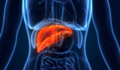
Liver Fibrosis Scan (Ultrasound Elastography) FibroScan Alternative
Liver fibrosis scanning in Leicester. Ultrasound elastography, blood tests, and a private GP review combined in one package.

Paediatric Ultrasound Scan Services
Private paediatric ultrasound scans in Leicester. Safe, radiation-free imaging for infants and children under expert consultant care.
Have a query about Ultrasound Scans – Weighing Up the Pros and Cons?
We recognise that getting the healthcare assistance you need can be difficult. So if you have a query, feel free to contact us and one of our treatment co-ordinators will be happy to help. We aim to reply to all queries within 24 hours (Mon – Fri).