Protecting muscle and bone during weight loss on GLP-1 medication
- Published:
- Last medically reviewed:
GLP-1 medications work. The weight comes off, often substantially, and for many people, that produces genuine improvements in metabolic health, joint pain, sleep and cardiovascular risk.
What is less discussed is what the weight is made of. Weight loss of any kind removes lean tissue alongside fat, and the faster the loss, the greater the proportion that is lean. On medication that produces large reductions in appetite, that proportion can be significant. This matters because muscle and bone are difficult to rebuild, particularly after 50, and because the scales cannot tell you what you are losing.
What the evidence shows
Across trials of GLP-1 and dual-agonist medications, a meaningful share of total weight lost is lean mass. The DXA substudy of SURMOUNT-1, in which 160 participants were scanned at baseline and again at 72 weeks, recorded reductions of 21.3% in body weight, 33.9% in fat mass and 10.9% in lean mass on tirzepatide, against 5.3%, 8.2% and 2.6%, respectively, on placebo [1]. The figures vary by study, population and how lean mass is measured, but the pattern is consistent.
Some of that lean loss is expected and appropriate. A larger body carries more muscle to move itself, and some reduction accompanies any weight loss, including surgical and dietary. The concern is not that lean mass falls; it is that it can fall further and faster than necessary, and that nobody is usually measuring it.
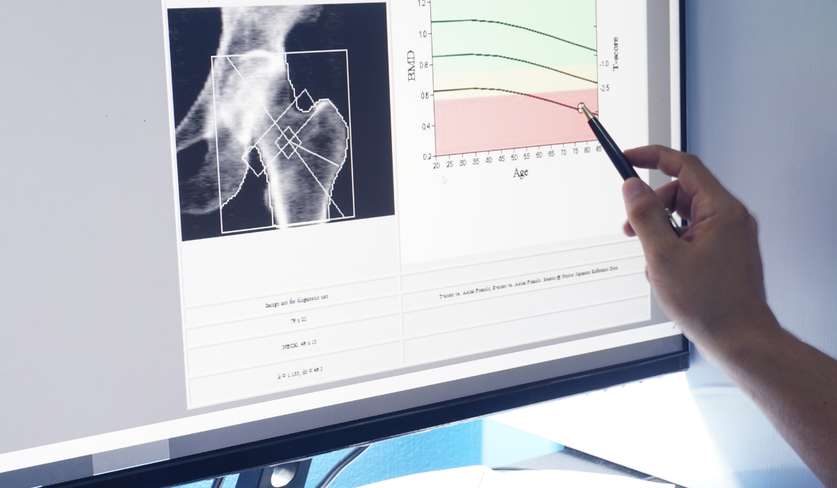
Bone is the less examined half of the question. Weight loss is associated with reductions in bone density, particularly at weight-bearing sites, and the mechanisms include reduced mechanical loading, altered hormonal signalling and lower intake of the nutrients bone requires. The long-term fracture implications of GLP-1 treatment specifically are not yet settled, which is a reason for measurement rather than a reason for reassurance.
Who is most exposed
- Adults over 50, in whom lean mass is already declining and the capacity to rebuild it is reduced.
- Post-menopausal women, who are losing bone for hormonal reasons at the same time.
- Anyone who was already low in muscle mass before starting — often people who have carried excess weight for a long time without resistance training.
- People losing weight very quickly, or with very low protein intake because appetite suppression has reduced overall food intake rather than reshaped it.
- Anyone with existing risk factors for osteoporosis, where an additional stimulus to bone loss is least welcome.
What helps
- Resistance training. The single most effective countermeasure. Two to three sessions a week working the major muscle groups substantially reduce lean mass loss during a calorie deficit and provide the loading stimulus bone needs. Walking is good for many things but does not do this job.
- Protein intake. Appetite suppression makes it easy to fall well below requirements, and protein is the first thing to drop when meals get smaller. Requirements during weight loss are higher, not lower, and spreading intake across meals improves the response. This usually needs planning rather than willpower.
- Rate of loss. Faster is not better. A more gradual reduction preserves more lean tissue, and dose titration can be adjusted with your prescriber if loss is running very quickly.
- Vitamin D, calcium and overall nutritional adequacy. Reduced food intake means reduced micronutrient intake unless it is attended to.
Measurement. All of the above is guesswork without knowing what is actually happening.
Why the scales cannot answer this
A person losing 12 kg has no way of knowing from the scales whether that was 10 kg of fat and 2 kg of lean tissue, or 8 kg and 4 kg. The number is identical. The outcome is not.
Body composition scanning separates them. A DEXA scan measures fat mass, lean mass by region, visceral fat and bone mineral content directly, with a repeatability of around 1 to 2% for fat mass — precise enough that a change over four to six months is real rather than noise.
Appendicular lean mass, the lean tissue in the arms and legs, is the specific measure used to assess sarcopenia [2], and it is the number worth watching through a period of substantial weight loss.
A sensible measurement approach
- Before starting, or as early as possible, a DEXA body composition scan to establish a baseline. If you have risk factors for bone loss, a bone density scan at the same time gives you a bone baseline too, and both can be done in one appointment.
- During treatment: bioimpedance every few weeks is enough to follow the direction of travel between scans, provided conditions are kept consistent.
- At four to six months: a repeat DEXA scan, compared against the baseline on the same machine, showing what proportion of the loss was fat and what was lean.
If lean loss is running high, that is actionable — protein, training and rate of loss can all be adjusted. Without measurement, it is invisible until it shows up as weakness or as a fracture years later.
Scanning at The Health Suite
We offer DEXA body composition and bone density scanning at our Leicester clinic, reported by a consultant radiologist, alongside our weight management service. Scans are available whether or not your treatment is with us.
DEXA Bone Density Scan
Precise DEXA bone density scanning in Leicester. Measure fracture risk, T-scores, and Trabecular Bone Score (TBS) with radiologist reporting.